Category: Lesion Expansion

Description

  • Evidence of progressive tissue loss in the core of chronic MS lesions: A longitudinal DTI study

    Evidence of progressive tissue loss in the core of chronic MS lesions: A longitudinal DTI study

    Progressive Tissue Loss in Chronic MS Lesions: Mechanisms of Brain Volume Loss

    This longitudinal study reveals how ongoing tissue damage within chronic MS lesions contributes to brain atrophy, offering new insights into disease progression even in treated patients.

    Key Findings:

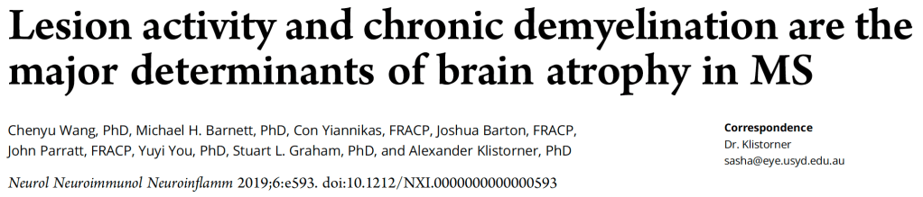
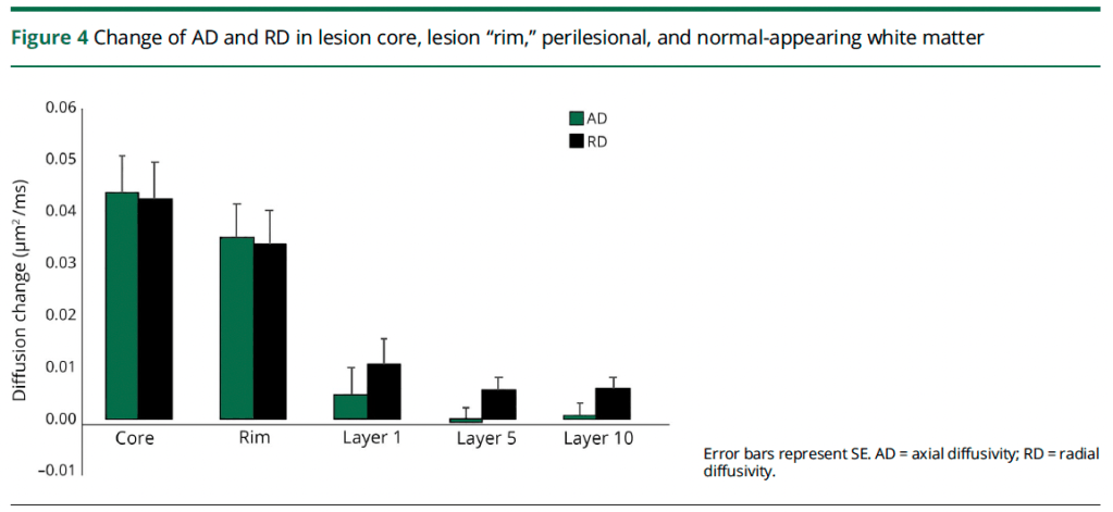
    • Significant tissue loss occurs within chronic inactive MS lesions, with water diffusion increasing by 3.3% annually in lesion cores
    • Brain volume decreased by approximately 2% over the 5-year study period, showing continued atrophy despite treatment
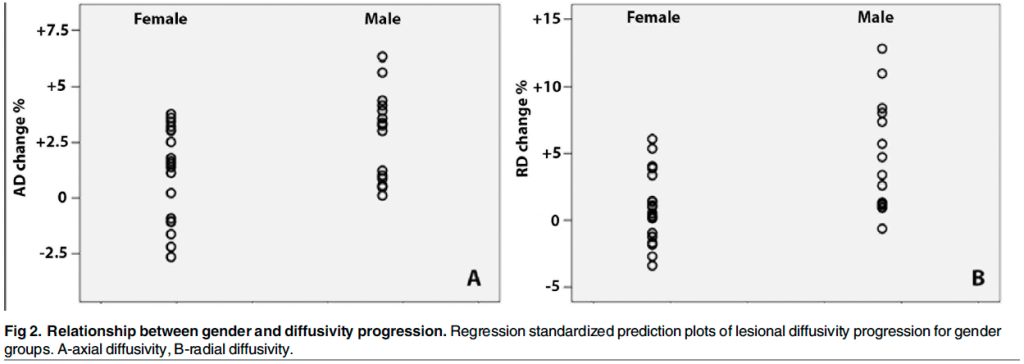
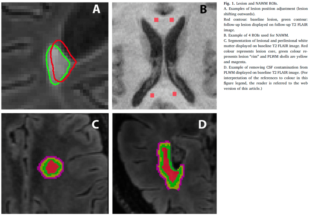
    • Males showed nearly twice the rate of tissue damage compared to females, suggesting important gender differences in disease progression
    • Tissue damage within chronic lesions strongly correlated with overall brain atrophy, explaining 38% of brain volume changes

    Novel Technical Advances:

    • Used advanced diffusion tensor imaging to track microstructural changes over time
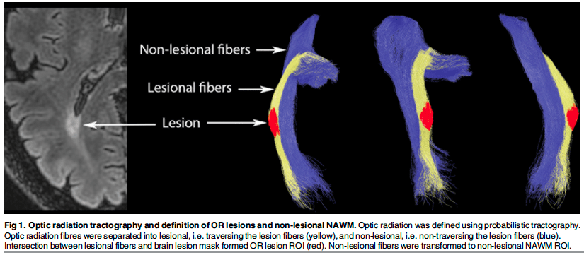
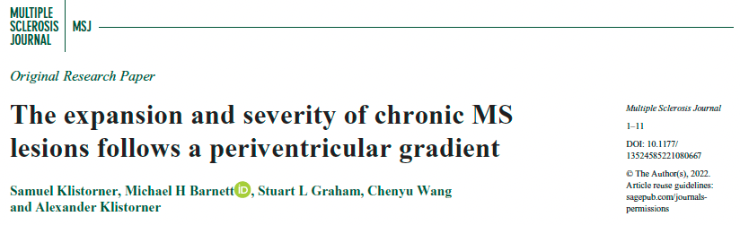
    • Developed methods to precisely analyze lesion cores separate from edges
    • Created techniques to account for brain atrophy when measuring lesion changes
    • Established ways to quantify progressive tissue damage in stable lesions

    Clinical Implications:

    • Explains why current MS treatments may not fully prevent disease progression
    • Suggests need for additional therapeutic approaches targeting chronic lesion damage
    • Highlights importance of considering gender differences in clinical trials
    • Demonstrates value of monitoring lesion changes to predict disease course

    Why It Matters:

    Understanding how chronic lesions continue to cause tissue damage even without new inflammation provides crucial insights into MS progression. The findings suggest that protecting demyelinated axons and promoting remyelination may be critical therapeutic goals, alongside current anti-inflammatory treatments.

  • Lesion activity and chronic demyelination are the major determinants of brain atrophy in MS

    Lesion activity and chronic demyelination are the major determinants of brain atrophy in MS

    Brain Atrophy in Multiple Sclerosis: Multiple Factors Drive Progressive Brain Loss

    This study reveals how different disease processes contribute to brain volume loss in multiple sclerosis (MS), providing new insights into why current treatments may not fully prevent disease progression.

    Key Findings:

    • Brain volume loss in MS results from multiple factors, with new lesions accounting for 40% of brain atrophy variability
    • Progressive tissue damage within chronic lesions significantly contributes to brain shrinkage, explaining 38% of brain volume changes
    • Diffuse damage to normal-appearing white matter adds another important factor driving brain tissue loss
    • The combination of these factors explains over 60% of brain volume changes in MS patients

    Novel Technical Insights:

    • Used advanced imaging to track tissue changes over 5 years in MS patients
    • Demonstrated that chronic lesions continue to cause damage long after their initial formation
    • Showed that damage to normal-appearing brain tissue occurs independently from lesion activity
    • Developed methods to measure different types of tissue injury simultaneously

    Clinical Implications:

    • Explains why current MS treatments, which mainly prevent new lesions, may not fully stop disease progression
    • Suggests need for combination therapies targeting multiple aspects of the disease
    • Provides new ways to monitor treatment effectiveness
    • Highlights importance of early intervention to prevent cumulative tissue damage

    Why It Matters:

    Understanding these multiple contributors to brain atrophy helps explain why MS progresses differently in different patients and points toward new therapeutic strategies. The findings suggest that effective MS treatment may require addressing both inflammatory activity and chronic tissue degeneration.

  • Expansion of chronic lesions is linked to disease progression in relapsing–remitting multiple sclerosis patients

    Expansion of chronic lesions is linked to disease progression in relapsing–remitting multiple sclerosis patients

    We investigated how chronic inflammatory lesions in multiple sclerosis evolve over time by studying their expansion patterns and relationship to disease progression in patients with relapsing-remitting MS over a 4-year period.

    Key Findings:

    • The expansion of chronic lesions, rather than formation of new lesions, accounts for most (67%) of total brain lesion volume increase in MS patients
    • Nearly half (46%) of chronic lesions showed significant expansion, while only 12% shrank over time
    • Lesion expansion strongly correlated with both brain atrophy and increased disability, suggesting it drives disease progression
    • Older patients showed higher rates of lesion expansion, potentially explaining accelerated disability in aging MS patients

    Novel Technical Advances:

    • Developed sophisticated imaging analysis methods to precisely track individual lesion changes over time
    • Created automated algorithms to distinguish between expanding chronic lesions and new lesion formation
    • Established quantitative measures to assess tissue damage within expanding lesions using diffusion imaging
    • Implemented rigorous controls for brain atrophy effects on lesion measurements

    Clinical Implications:

    • The dominant role of chronic lesion expansion in disease progression suggests new therapeutic targets are needed
    • Treatments focusing only on preventing new lesions may not adequately address ongoing tissue damage
    • Monitoring chronic lesion expansion could help identify patients at higher risk of progression
    • Age-related increases in lesion expansion indicate older patients may need different therapeutic approaches

    Why It Matters:

    This study fundamentally changes our understanding of how MS progresses by showing that the slow expansion of existing lesions, rather than formation of new ones, is the primary driver of accumulating disability. This challenges the traditional focus on preventing new lesion formation and suggests that therapies targeting chronic inflammation at lesion edges could help prevent disease progression. The findings explain why current treatments, while effective at reducing new lesions, may not fully prevent advancing disability, especially in older patients. This work provides crucial evidence for developing new therapeutic strategies aimed at the smoldering inflammation that causes lesion expansion.

  • Expansion of chronic MS lesions is associated with an increase of radial diffusivity in periplaque white matter

    Expansion of chronic MS lesions is associated with an increase of radial diffusivity in periplaque white matter

    Demyelination as a Key Driver in Multiple Sclerosis Lesion Expansion

    Using our in-house single-streamline diffusion tensor imaging analysis, we investigated the relationship between chronic lesion expansion and tissue damage patterns in relapsing-remitting multiple sclerosis (RRMS), focusing on the role of demyelination and axonal injury.

    Key Findings:

    • Sequential Tissue Damage: Our analysis revealed that demyelination likely represents the initial step in chronic lesion expansion, preceding significant axonal damage
    • Differential Impact: We found that the rate of myelin damage in expanding lesions was twice that of axonal injury in surrounding tissue, suggesting demyelination as the primary early mechanism
    • Predictive Markers: Higher levels of myelin damage in periplaque white matter strongly predicted subsequent lesion expansion, while axonal damage markers showed weaker correlation
    • Expanding vs Stable Lesions: Expanding lesions showed significantly higher rates of ongoing demyelination compared to stable lesions, particularly in periplaque areas

    Clinical Implications:

    • These findings suggest that targeting early demyelination could be crucial for preventing lesion expansion
    • The identification of demyelination as an initial step provides a potential window for therapeutic intervention
    • Our results support the development of remyelinating therapies as a strategy to prevent associated neurodegeneration

    Why It Matters:

    Understanding the sequence of tissue damage in expanding MS lesions helps identify optimal timing for therapeutic interventions. By demonstrating that demyelination precedes significant axonal loss, this research suggests that early treatment targeting myelin protection or repair could help prevent the cascade of tissue damage that leads to progressive disability.

  • Examining the Relative Contribution of Slow-Burning Inflammation and Chronic Demyelination to Axonal Damage in Chronic Multiple Sclerosis Lesions

    Examining the Relative Contribution of Slow-Burning Inflammation and Chronic Demyelination to Axonal Damage in Chronic Multiple Sclerosis Lesions

    Our study compared how two different mechanisms – slow-burning inflammation at lesion edges and chronic demyelination – contribute to tissue damage in multiple sclerosis lesions.

    Key Findings:

    • Expanding vs Stable Lesions: Of 361 lesions analyzed, 104 showed expansion while 257 remained stable, demonstrating distinct patterns of tissue damage
    • Inflammation Impact: Expanding lesions showed 5 times higher tissue damage rate compared to stable lesions, indicating slow-burning inflammation’s significant role
    • Demyelination Effects: Analysis of chronically demyelinated optic nerves revealed modest tissue damage similar to stable brain lesions, suggesting limited impact of demyelination alone

    Novel Technical Advances:

    • Developed method to normalize measurements of tissue damage across lesions
    • Created approach to exclude effects of new lesions on chronic lesion analysis
    • Used visual system to validate findings in a different part of the nervous system

    Clinical Implications:

    • Slow-burning inflammation appears more destructive than chronic demyelination
    • Treatments targeting inflammation at lesion edges may be more effective than those focusing on demyelination
    • Findings suggest need for early intervention to prevent lesion expansion

    Why It Matters:

    Understanding the relative impact of these two mechanisms helps guide treatment strategies for MS. Our findings suggest that controlling slow-burning inflammation at lesion edges should be a primary therapeutic target, potentially leading to more effective treatments that could slow disease progression.

  • Differentiating axonal loss and demyelination in chronic MS lesions: A novel approach using single streamline diffusivity analysis

    Differentiating axonal loss and demyelination in chronic MS lesions: A novel approach using single streamline diffusivity analysis

    We developed an innovative method to analyze how multiple sclerosis lesions evolve over time, allowing us to separately measure and track nerve fiber loss and myelin damage using advanced brain imaging techniques. This new approach provides crucial insights into how MS causes progressive disability.

    Key Findings:

    • Our analysis revealed that nerve fiber loss and myelin damage contribute independently to MS lesion development, with different patterns of change over time
    • Nerve fiber loss shows a direct relationship with lesion severity – as lesions worsen, more nerve fibers are lost in a predictable way
    • Myelin damage has the greatest impact in early lesions when many nerve fibers are still intact, but becomes less prominent as nerve fiber loss increases
    • The rim area around lesions shows distinct patterns from the lesion core, making it potentially useful for monitoring treatment effects

    Novel Technical Advances:

    • Created a new single-fiber tracking method that can analyze MS lesions throughout the brain, not just in specific nerve pathways
    • Developed algorithms to separate and quantify nerve fiber loss from myelin damage within the same lesion
    • Established techniques to control for complex brain anatomy when measuring lesion changes
    • Implemented automated analysis approaches that enable consistent measurement across many patients

    Clinical Implications:

    • Provides a way to identify which lesions may respond best to remyelination therapies
    • Enables better monitoring of treatment effects by separately tracking nerve fiber and myelin changes
    • Suggests the lesion rim may be the optimal target for treatments aimed at protecting nerve fibers
    • Demonstrates why current MS treatments may have limited effects on progressive disability

    Why It Matters:

    This work fundamentally changes our understanding of how MS lesions evolve and cause disability. By showing that nerve fiber loss and myelin damage follow distinct patterns, we can better understand why some treatments work better than others and identify new therapeutic strategies. The ability to separately measure these two processes provides critical tools for developing and testing new treatments, particularly those aimed at protecting nerve fibers or promoting myelin repair. This advance may help explain why some patients progress faster than others and guide more personalized treatment approaches in MS.

  • Quantifying Chronic Lesion Expansion in Multiple Sclerosis: Exploring Imaging Markers for Longitudinal Assessment

    Quantifying Chronic Lesion Expansion in Multiple Sclerosis: Exploring Imaging Markers for Longitudinal Assessment

    Our study investigated methods to accurately measure and predict chronic lesion expansion in multiple sclerosis (MS) using imaging data collected over 1-2 years, focusing on markers that don’t require longitudinal lesion segmentation.

    Key Findings:

    • Central Brain Atrophy (CBA): Measured over 2 years, CBA accurately predicts 4-year lesion expansion with 94% sensitivity and 85% specificity
    • Mean Diffusivity (MD): Changes in MD within chronic lesions strongly correlate with future expansion, providing 81% sensitivity and specificity
    • Progressive Volume/Severity Index: We developed this novel measure combining lesion volume changes and tissue damage severity to standardize assessment across patients

    Clinical Implications:

    • CBA offers a reliable early indicator of future lesion expansion without requiring complex lesion segmentation
    • The combination of multiple markers provides comprehensive assessment of disease progression
    • These findings enable shorter study periods for clinical trials evaluating new treatments

    Why It Matters:

    Early identification of patients likely to develop expanding lesions could inform treatment decisions and improve outcomes. Our markers provide practical tools for monitoring disease activity in clinical settings and trials.

  • Progressive Injury in Chronic Multiple Sclerosis Lesions Is Gender-Specific: A DTI Study

    Progressive Injury in Chronic Multiple Sclerosis Lesions Is Gender-Specific: A DTI Study

    Gender-Specific Progression of Tissue Damage in Multiple Sclerosis Lesions

    Using our in-house DTI analysis techniques, we investigated how chronic MS lesions evolve differently in male and female patients, uncovering important insights into gender-specific disease progression patterns.

    Key Findings:

    • Gender-Specific Evolution: Male patients showed significantly faster progression of tissue damage in chronic lesions, with up to 6.5 times higher rates of change in certain markers compared to females
    • Comprehensive Changes: We observed increases in both axial and radial diffusivity within lesions, indicating ongoing inflammation and tissue destruction that particularly affected male patients
    • Distinct Patterns: While lesion size was larger in males, statistical analysis confirmed that gender itself, rather than lesion volume, drove the differences in progression

    Clinical Implications:

    • These findings help explain the observed trend of faster clinical progression in male MS patients
    • The results establish DTI as a promising biomarker for monitoring gender-specific disease evolution
    • The data suggests potential need for gender-specific treatment approaches

    Why It Matters:

    Understanding gender-based differences in MS progression provides crucial insights for developing more personalized treatment strategies. This research offers the first clear biological evidence explaining why male patients often experience faster disease progression, potentially leading to more targeted therapeutic interventions based on gender-specific disease patterns.

  • Diffusivity in multiple sclerosis lesions: At the cutting edge?

    Diffusivity in multiple sclerosis lesions: At the cutting edge?

    Patterns of Tissue Damage in Multiple Sclerosis Lesions: Insights from Advanced Diffusion Imaging

    Using our in-house diffusion tensor imaging (DTI) analysis techniques, we investigated how tissue damage evolves differently in the core versus periphery of chronic MS lesions, uncovering distinct patterns that provide new insights into disease mechanisms.

    Key Findings:

    • Distinct Damage Patterns: We identified a previously unrecognized pattern where lesion rims show predominantly myelin damage, while core regions exhibit more severe tissue destruction and axonal loss
    • Rim Characteristics: The peripheral regions of lesions maintain relatively constant width regardless of lesion size, suggesting a preserved zone that may be more amenable to therapeutic intervention
    • Core Dynamics: Central lesion areas demonstrate more severe tissue disruption with equal increases in both axial and radial diffusivity, indicating extensive structural breakdown

    Clinical Implications:

    • The preserved lesion rim may represent an optimal target for emerging remyelination therapies
    • Understanding these distinct patterns could help develop more sensitive measures for monitoring treatment effects
    • The findings suggest different therapeutic approaches may be needed for lesion cores versus rims

    Why It Matters:

    This research reveals for the first time how tissue damage evolves differently across MS lesions. By demonstrating that the lesion rim maintains structural integrity while showing primarily myelin loss, we’ve identified a potential therapeutic window where remyelination treatments might be most effective. This insight could fundamentally change how we approach treatment and monitoring of MS lesions.

  • The expansion and severity of chronic MS lesions follows a periventricular gradient

    The expansion and severity of chronic MS lesions follows a periventricular gradient

    Using our in-house LEAP methodology, we analyzed how the proximity of chronic lesions to cerebrospinal fluid (CSF) spaces affects their expansion and severity in patients with relapsing-remitting multiple sclerosis (RRMS).

    Key Findings:

    • Periventricular Gradient: Lesions closer to the ventricles exhibit faster expansion and more severe tissue damage compared to those further away.
    • Progressive Axonal Damage: Periventricular lesions showed higher rates of axonal loss and microstructural damage, driven by slow-burning inflammation at the lesion rim.
    • CSF-Related Dependency: The proximity to CSF appears to amplify cytotoxic factors that promote lesion expansion and tissue injury.

    Clinical Implications:

    • These findings highlight the importance of monitoring periventricular lesion dynamics as a marker for MS progression.
    • The CSF-related gradient offers potential insights into the underlying mechanisms of chronic inflammation in MS and may inform targeted therapeutic strategies.

    Why It Matters:

    Understanding the spatial and biological factors driving lesion expansion enables better tracking of disease activity and could guide more personalised treatment approaches for MS patients.